Mesapamea |
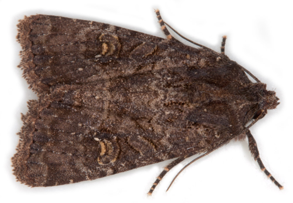
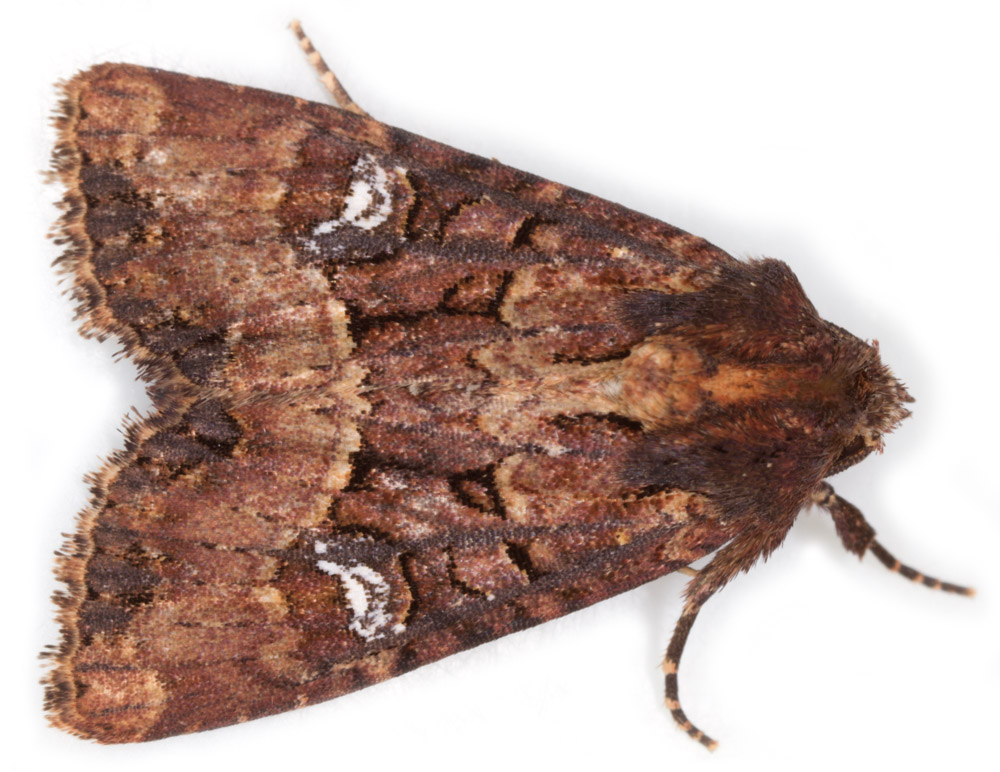
These two species are very similar and both show a wide range of variation in forewing colour and markings. The two species were not separated until 1983 at which time a third species, M.remmi (Remm's Rustic), showing intermediate genital features, was also accepted - but this is now thought to be a hybrid. M.secalis/M.didyma can only be separated on genital features.
Ref: Difficult Species Guide
Ref: Difficult Species Guide
Males
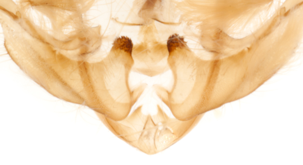
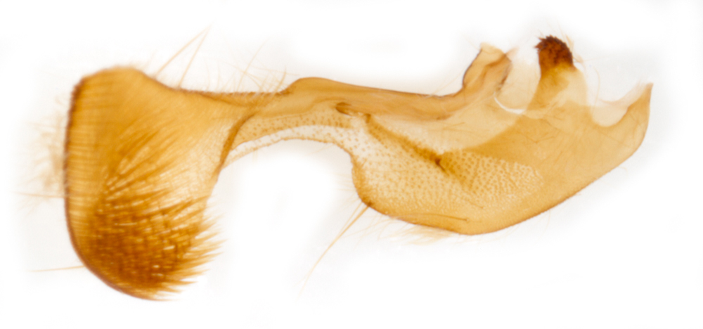
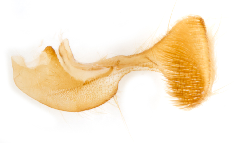
Clavus (a projection from the base of the costa of the sacculus = base of the dorsal margin of the valva as a whole)
In M.secalis it is heavily sclerotised, protrudes to a greater extent (in a dorsal direction?) and has "many short teeth" at the apex.
In M.didyma it is weakly sclerotised, protruded to a lesser extent (in a more medial direction?) and has "fine setae".
Cornutus
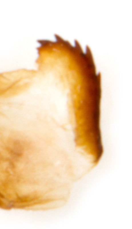
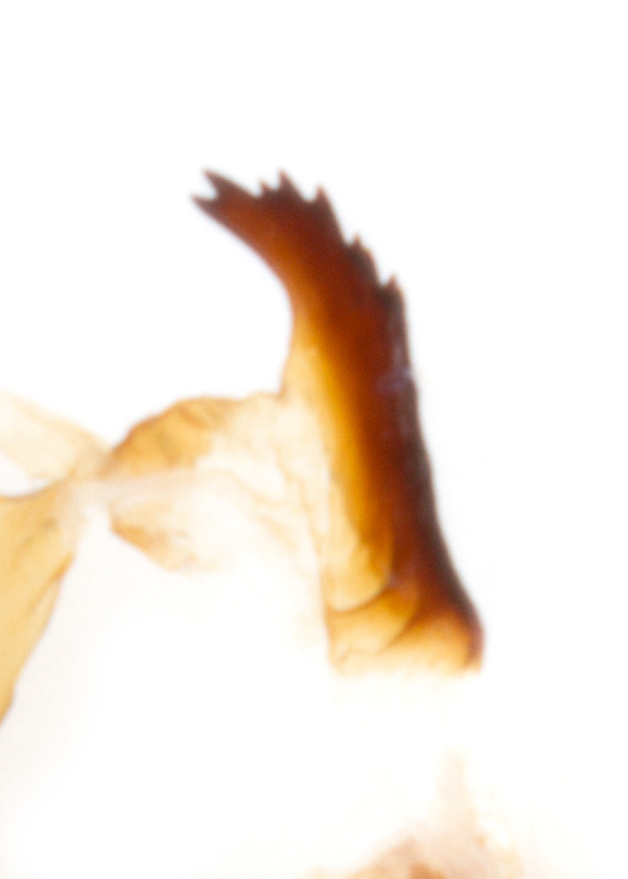
Both species have a single moderately large cornutus attached along one edge to the vesica near the apex of the aedeagus, and free along its other edge, which is dentate in its apical half. In M.secalis the cornutus is broad and rounded and the attachment to the vesica extends to its dentate apex; in M.didyma the cornutus is long and narrow and the attachment to the vesica leaves the apical ~2/5 free, such that there is a smooth (non-dentate) margin between the attachment to the vesica and the apex of the cornutus.
Both species have a single moderately large cornutus attached along one edge to the vesica near the apex of the aedeagus, and free along its other edge, which is dentate in its apical half. In M.secalis the cornutus is broad and rounded and the attachment to the vesica extends to its dentate apex; in M.didyma the cornutus is long and narrow and the attachment to the vesica leaves the apical ~2/5 free, such that there is a smooth (non-dentate) margin between the attachment to the vesica and the apex of the cornutus.
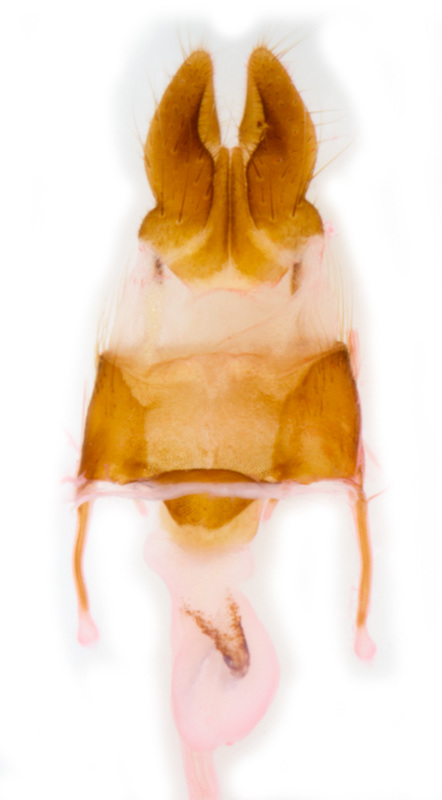
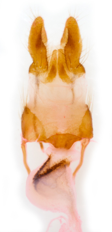
Females
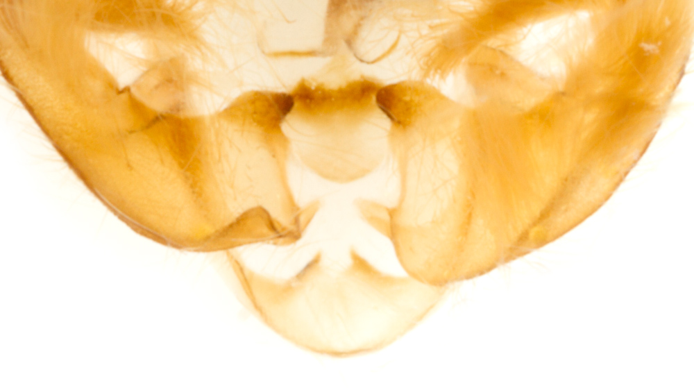
In both species there is a swelling with some internal sclerotisation at the posterior end of the ductus bursae. In the standard ventral view, with the ovipositor at the top, this swelling is to the right in M.secalis and to the left in M.didyma. The sclerotisation also seems to be heavier in M.didyma.
In both species there is a swelling with some internal sclerotisation at the posterior end of the ductus bursae. In the standard ventral view, with the ovipositor at the top, this swelling is to the right in M.secalis and to the left in M.didyma. The sclerotisation also seems to be heavier in M.didyma.
