Nemapogon identification
Nemapogon is a difficult genus to confirm specific identity.
N.clematella is distinctive being basically white with an angulate blackish median fascia.
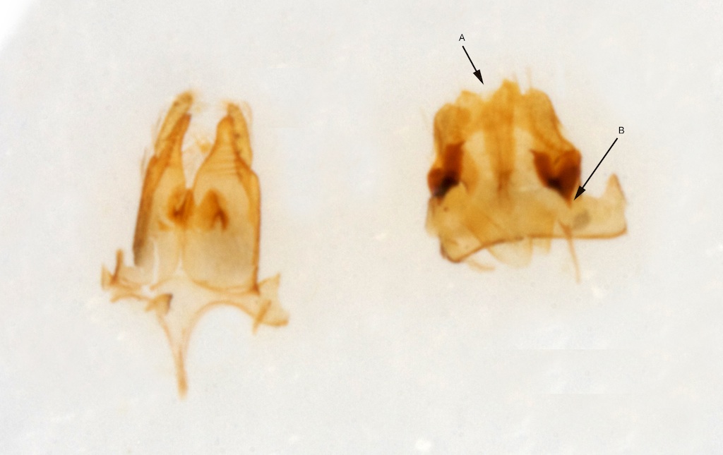
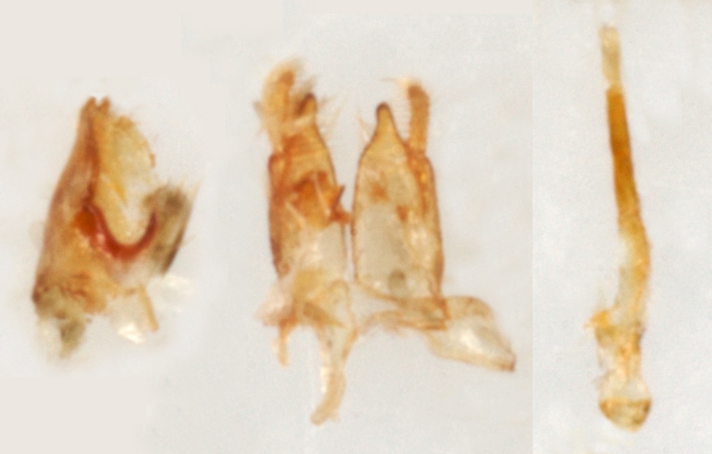
The remaining species are similar and variable and may require genital examination to confirm ID.
N.inconditella, N.varietella, N.ruricolella and N.picarella tend to have white heads.
N.inconditella has only occured once in UK.
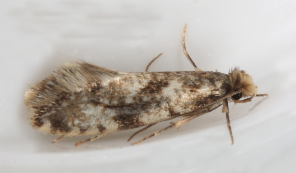
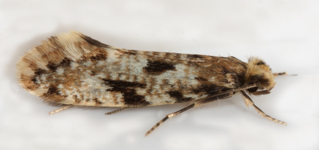
N.granella, N.cloacella and N.koenigi all have ochreous heads.
According to MBGBI2 N.granella is the only species in this group that lacks a clear white postmedian spot in the disc of forewing, but I have found the presence of this spot not easy to identify with certainty in individuals I am pretty sure are N.cloacella or N.koenigi.
Forewing ground colour is more uniformly dark in both N.granella and N.koenigi than in N.cloacella.
Where N.cloacella and N.koenigi appear similar they can usually be separated by the dark median costal blotch which is more clearly defined in N.cloacella and is lost in the darker ground colour below (dorsal to) the costa in N.koenigi.
Where N.cloacella and N.ruricolella appear similar they can usually be distinguished by the more uniform reddish ochreous ground colour of the latter especially in the apical area of the forewing.